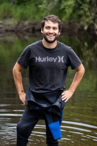
VIMS
Miguel Montalvo homepage

Miguel Montalvo

Ph.D. Student

Email: [[v|mmontalvo]]
Phone: 804-684-7544
Office: Fisheries Science Laboratory 110
Advisor: {{https://www.vims.edu/about/directory/faculty/hilton_ej.php, Dr. Eric Hilton}}

About me

As a person whose background is in marketing and communications, I strive to leave my mark in the world of science by using visual media to get people excited about the natural world. I often use microphotography to capture small study animals and their details either as stills or on film to showcase my work. I also enjoy wildlife photography and videography and I create content that is often featured in the social media platforms of different scientific organizations (VIMS, The Smithsonian National Museum of Natural History, Mote Marine Laboratory and Aquarium, etc).

My work at VIMS is centered around morphology and systematics of fishes, which means that I am looking at their body features (bones, muscles, etc) and trying to define their evolutionary relationships. I am passionate about this work because I get to work with real specimens and use techniques that are often visually striking (see some of the images below).

Current projects
Billfishes and fishes with a bill

I am investigating the evolution and development of the billfishes (the group of fishes that includes Marlins, Sailfish and Swordfish). These animals are fascinating because, among other things, they have an elongated rostrum (a bill), they are very fast swimmers, are really fast growers, and some can reach enormous sizes—like the Atlantic Blue Marlin (Makaira nigricans) in the following photo.

This is me at a fishing tournament with an enormous Atlantic Blue Marlin - this was my first time seeing a fish of this size! We collect data from tournaments like this to help us assess the health of fish stocks. Some of these fishes are sometimes donated to us and become part of the Nunnally Ichthyology Collection that houses specimens that can be used by researchers all over the world.

This is me at a fishing tournament with an enormous Atlantic Blue Marlin - this was my first time seeing a fish of this size! We collect data from tournaments like this to help us assess the health of fish stocks. Some of these fishes are sometimes donated to us and become part of the Nunnally Ichthyology Collection that houses specimens that can be used by researchers all over the world.

As part of this project, I will also be looking at the larval stages of these fishes to study their development (the field of biology that studies the development of organisms is called ontogeny, by the way). It is amazing to think that a fish as large as the one in the photo starts as a larva a few millimeters in length (see the following photo of a cleared and stained Swordfish larva). Studying the ontogeny of these animals can give us clues as to some of their evolutionary relationships. Another component of this project would be to look at fossils related to these remarkable creatures. I am especially looking forward to this because I love fossils!

Cleared and stained image of a Swordfish larva. This larva is around 10mm in total length. The bones are stained in red and the cartilage is blue.

This the larva of a Swordfish (Xiphias gladius). It has been cleared and stained to show the bony and cartilaginous features.

Lastly, there are other fishes that have elongated rostra (like needlefishes and cornetfishes) and there may be surprising connections between these diverse groups of fishes whose commonality is the rostral feature. I will be using techniques such as clearing and staining (see below), CT scanning, and specimen dissections to try to find more about why some of these fishes evolved such morphological characters, and how they differ from one another.

This is a sample of a fish that I have cleared and stained, and then photographed it through the microscope. This technique allows you to see the bones and cartilage of the fish unobscured by muscles and other tissues. The bones are stained in red and the cartilage is stained in blue. This is a Butterfish (genus Peprilus) that I dissected after clearing and staining, and I arranged in a sort of exploded view.
Larval fishes

Baby fishes are fascinating, they are tiny, and derpy, and they—most of the time—look nothing like their adult parents. As it happens, VIMS has a long legacy of larval fishes research which makes it the perfect place to get more acquainted with these amazing animals. So, after pestering some of my colleagues who are actively researching them, I am helping them to sort and document some larval specimens.

Because these specimens are so tiny, you use a stereo microscope to look and sort through the samples. I am using my own Leica scope, with a phototube attached to a my Nikon mirrorless to take images - and sometimes videos. I hope you are as captivated by these specimens as I am :)

Pelican gulper eel larva

Closeup image of the mouth

Lefteye flounder larva (Bothus sp.)Larval Anglerfish - Gigantactis sp.Striped Scolar (Diplospinus multistriatus) larva

Interests

Fishes, fossils, conservation, sustainability, field science, science communications and marketing, diving, photography, birdwatching.