

Taxonomy of Hematodinium
TAKEN FROM STENTIFORD & SHIELDS, 2005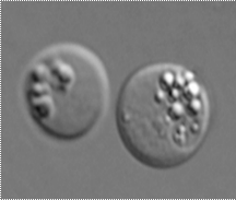
Members of the genus Hematodinium are primarily parasites of decapod crustaceans. They can be identified as dinoflagellates on the basis of their typical dinokaryon (or mesokaryon, Chatton 1920), the alveolate pellicle, the presence of naked, athecate gymnodinoid dinospores (or zoospores), and the classic form of mitosis known as dinomitosis (similar to that reported for the genus Syndinium, see Ris & Kublai 1974). Due to their lack of distinct characters and their poorly understood life cycles, there are only two described species of Hematodinium. The type species, Hematodinium perezi, was first described from the portunid crabs Carcinus maenas and Liocarcinus depurator off Luc-sur-Mer and Roscoff, on the coast of Normandy, France, and off Banyuls-sur-Mer, on the Mediterranean coast of France (Chatton & Poisson 1931). Unfortunately, Chatton and Poisson (1931) may have selected the wrong hosts for study because at the time, only 3 of 470 L. depurator and a few of 3000 C. maenas were infected. Since then, Hematodinium perezi, or a closely related species, has been reported in epidemics from Cancer pagurus (Latrouite et al. 1988; Stentiford et al. 2002) and Necora puber (Wilhelm & Boulo 1988, Wilhelm & Mialhe 1996) off Brittany, France, and from the English Channel (Stentiford et al. 2003). A second species, H. australis, was described from Australia (Hudson & Shields 1994). It was separated from H. perezi on the basis of size of the vegetative stage (trophont), the presence of rounded plasmodial stages and the austral location. Molecular studies later supported the separation of H. australis from other known forms of Hematodinium (Hudson & Adlard 1996), but to date there have been no molecular or ultrastructural studies on the type species from the type host. The Hematodinium parasite from N. norvegicus (Field et al. 1992) and that from C. bairdi (Meyers et al. 1987) are no doubt new species, but until there is comparative work with the type species, it will remain difficult to place them within the genus. This lack of comparative study with the type species has hampered the taxonomy of the group. At present, there are only a few characters that can be used to distinguish among species. The presence in a crustacean host, the hemolymph-dwelling filamentous plasmodial stages, and the high intensity infections with amoeboid trophonts and their ever present dinokaryon lead to the diagnosis of Hematodinium. The presence of the motile vermiform plasmodium, or filamentous trophont of Appleton and Vickerman (1998), may be of significance in the taxonomy of the genus as it only occurs in C. maenas (Chatton & Poisson 1931), C. sapidus (Newman & Johnson 1975; Messick 1994; Shields & Squyars 2000) and N. norvegicus (Field et al. 1992). It has not been reported for infections in C. bairdi (Meyers et al. 1987; Eaton et al. 1991), but a filamentous form has been observed in tissue preparations of C. opilio from Newfoundland (J. Shields, unpublished data).
DNA-based molecular diagnostics techniques appear to be particularly applicable to Hematodinium. Nucleotide sequences of a region of the small subunit rDNA (ssu rDNA) have been identified from several isolates of Hematodinium from a number of decapod species (Hudson & Adlard 1994, 1996, Bower et al. 2003); and that from Hematodinium sp. isolated from C. sapidus is available at GenBank (accession no. AF421184 and AF286023) (Gruebl et al. 2002). While their study did not include the type genus and species (H. perezi ex C. maenas), Hudson & Adlard (1996) found substantial sequence variation among the different species of Hematodinium from N. norvegicus, C. bairdi and C. sapidus . However, although recent studies have shown that parasites detected in other crab species (such as C. tanneri) are indeed species of Hematodinium, for further definition to species level using molecular-based techniques, other regions of the genome (such as the ITS region of the rDNA) must be analysed (Bower et al. 2003). That is, the existing DNA probes used for the detection of Hematodinium appear to be genus specific and not species specific (Shields, pers. observation). However, PCR primer sets are useful for diagnosing Hematodinium and have been used for verification of infections (Gruebl et al. 2002, Stentiford et al. 2002, Sheppard et al. 2003).