

Diagnosis of Hematodinium
Hematodinium infections can be diagnosed several different ways including:
Visual Examination
Wet Smears
Neutral red staining
Histology
Molecular detection
See below for examples of each method
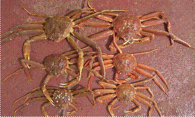
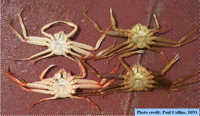
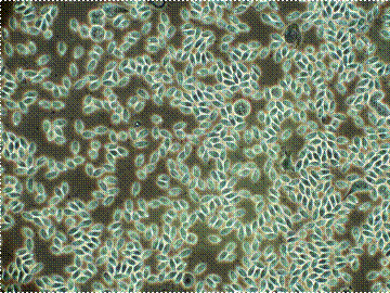
Visual Examination
|
|
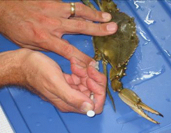
Wet Smears
|
|
|
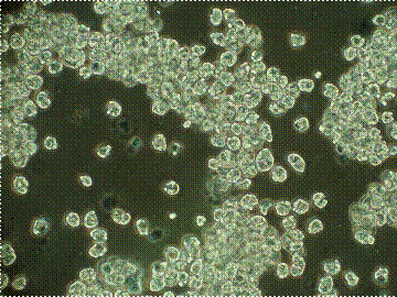
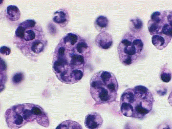
Neutral Red
 |
 |
Staining by neutral red is probably the easiest way for those who don't work with the parasite. However, the parasite may not take up the stain when cultured in long-term cultures. Ameboid trophonts (left) and host granulocytes (center) stained with neutral red (0.05% in saline buffer). Photographs are at different magnifications. Hemocytes often take up very little stain, but Hematodinium parasites rapidly take it up. Plasmodial form of the parasite in action stained with neutral red (right).
|
|
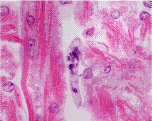
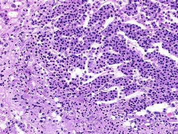
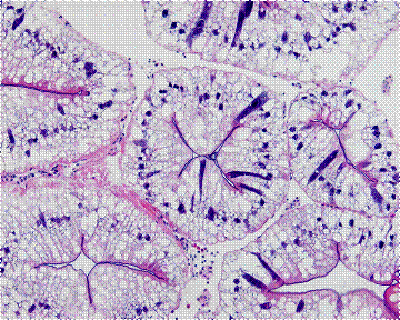
Histology
Hematodinium is like other dinoflagellates. The chromatin is condensed within the nucleus. Nuclei appear as if in arrested metaphase. Note the deeply blue-black staining chromatin within the nuclei of the parasites. These slides were staind with Hematoxylin and Eosin.
|
|
|
|
|
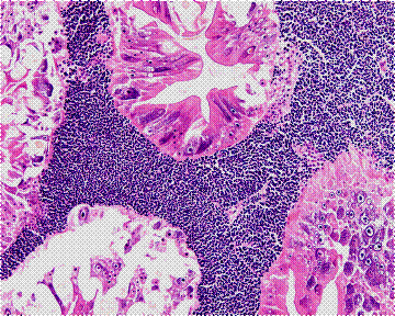 |
Hepatopancreas fron uninfected (left) and infected (right) snow crabs. The heavy infection results in a loss of connective tissue, a loss of hemocytes, and an enlarged hemal space between the tubules. |
|
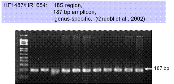
Molecular Detection
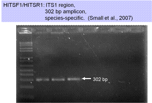 |
Routine diagnosis of Hematodinium:
|
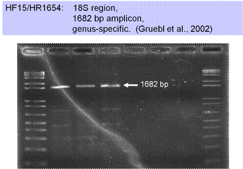 |
Primers targeting 18S region will be used to identify potential different strains of Hematodinium in blue crab. |
 |
Hematodinium quantification in water column:
|